Here's an essential for management of the postoperative patient...
Where does PE originate from?Typically after propogation and embolism of thrombi that develop in deep veins... 79% of patients with PE have evidence of calf DVT.
What are pathophysiologic sequelae of PE?
1. Anatomic obstruction of pulmonary arteries... although pulmonary infarction is typically avoided since lungs are also supplied by bronchial arteries.
2. Platelets cause the release of serotonin and vasoactive substances resulting in elevated pulmonary vascular resistance and V/Q mismatch.
3. RV afterload increases, leading to RV dilatation, ischemia, and right sided heart failure.
What are chronic sequelae of DVT and PE?
Post-thrombotic syndrome (chronic leg pain/swelling)
Chronic thromboembolic pulmonary hypertension
What are classic risk factors for DVT?
Virchow's triad - stasis, injury, and hypercoagulability
Also advanced age, obesity, pregnancy + postpartum period, malignancy, reduced mobility, polycythemia vera, smoking, central venous catheters, HITS, antiphospholipid antibodies, lupus anticoagulant
Medications - chemotherapy, oral contraceptives (3X risk), hormone-replacement therapy (2x risk), tamoxifen
Acutely-ill, hospitalized, medical patients have a risk of 15%
What specific populations are at high risk for DVT?
Total hip and knee replacement (risk of >50% without prophylaxis)
Cancer surgery
Major trauma
Spinal cord injury
What are genetic disorders that predispose to risk of venous thromboembolism?
Protein C and S deficiency
Antithrombin deficiency
Factor V Leiden
Prothrombin mutation (g20210a)
Hyperhomocyteinemia (C677T mutation)
Dysfibrinogenemia
Plasminogen deficiency
What isthe most common genetic risk factor for thrombophilia?
Factor V Leiden. This causes resistance to activated protein C
What are signs and symptoms of lower extremity DVT?
Leg pain, tenderness, warmth, or swelling
Homan's sign - calf pain with passive dorsiflexion at the ankle
What are signs and symptoms of PE?
dyspnea (73%)
tachypnea (70%)
pleuritic chest pain (66%)
rales / crackles (51%)
cough (37%)
Tachycardia (30%)
Leg swelling (28%) or pain (26%)
Fourth hearth sound (24%)
Loud P2 (23%)
Hemoptysis (13%)
Palpitations (11%)
JVD, systolic murmur, right sided gallop
syncope
hypotension
hypoxemia
PEA/cardiac arrest
Cardiogenic shock is diagnosed in 5% of acute PE.
While hypoxia and an Aa gradient is common, a normal ABG is seen in up to 20% of PE.
What are ECG findings of PE?
Tachycardia
V1-V4 T wave inversion
S1-Q3-T3 pattern
Right bundle branch block
P-wave pulmonale
Right axis deviation
These are nonspecific and are more often seen with very large PE
What are abnormal findings on CXR attributed to PE?
Westermark's sign - focal peripheral oligemia caused by PE obstruction
Hampton's hump - peripheral wedge seen above diaphragm
Palla's sign - enlarged right descending PA
How does D-dimer testing help with a diagnosis of PE?
This is a sensitive (96-98%) but very non-specific test.
It must be considered together with clinical suspicion and is better for patients presenting to the emergency room than for general surgical postop patients.
If the DDimer is negative in patients with a low-to-moderate pretest probability, imaging is not useful since PE is very unlikely.
If the patient has a high pretest probability, don't bother with checking a DDimer and go straight to imaging.
What imaging strategies are used for diagnosis of PE?
Spiral CT-angiography (90% sensitivity, 95% specificity)
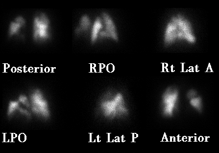
Ventilation/Perfusion Scanning - limitation: frequently non-diagnostic
Pulmonary arteriogram
Venous duplex for DVT
Echocardiography for hemodynamically significant PE
Use caution when Cr>1.5 with CTA
Avoid V-Q scanning if there is associated cardiopulmonary disease noted on CXR
What are treatment stategies for DVT?
Outpatient management with low-molecular weight heparin transitioning to warfarin with a goal INR of 2-3 for 3-6 months
Pregnant patients can't take warfarin and must be kept on a LMWH...
What are treatment strategies for PE?
PE should be managed by inpatient hospitalization.
Parenteral unfractionated heparin (therapeutic PTT), low-molecular weight heparin, or fondaparinux for at least 5 days while starting warfarin - bridging therapeutic INR of 2-3 for 2 consecutive days, continuing treatment for 3-6 months.
Factor VII - measured by the INR - has a half life of 6hrs... however thrombin (factor II) depletion takes 5 days...
How is unfractionated heparin administered?
It is bolused 60-80 U/kg, followed by a continuous infusion of 18 U/kg/hr. The infusion is titrated to a therapeutic PTT (60-80 seconds) by checking the PTT level every 6 hrs.
What are benefits of LMWH?
These have more predictible pharmacokinetics and greater bioavailability than unfractionated heparins, allowing subcutaneous administration once or twice per day.
Therapeutic dosing for enoxaparin is 1mg/kg SC BID or 1.5mg/kg SC QD
How can you monitor if levels of LMWH are therapeutic?
Check anti-factor Xa levels
This is not as convenient as a PTT, but consider this in morbidly obese or pregnant patients, or if there have been issues with renal function.
What is fondaparinux?
A synthetic pentasaccharide that inhibits Factor Xa
Structurally, it looks like the antithrombin-binding portion of heparin. It is administered once daily, subcutaneously.
This should not be used in patients with severe renal insufficiency.
What if the patient has heparin-induced thrombocytopenia with thombosis?
The patient should be treated with direct thrombin inhibitors (argatroban or lepirudin). Warfarin treatment should be held until the disease process is controlled and the platelet count has normalized to avoid thrombotic complitcations and warfarin-induced skin necrosis.
Lepirudin is excreted by the kidneys and argatroban is metabolized by liver.
What is the risk of death from recurrent thromboembolism in patients treated for DVT?
0.4%
What is the risk of death from recurrent thromboembolism in patients treated for PE?
1.5%
What are indications for a vena caval filter?
Contraindications to anticoagulation
Major bleeding complications during anticoagulation
Recurrent DVT or PE while receiving adequate therapy
Retrievable filters can often be inserted... although often times they are forgotten to be removed!
The tide is beginning to turn against prophylactically placing IVC filters, but they may be recommended in select trauma patients.
When is thrombolytic therapy considered for management of PE?
If the PE is severe enough to cause cardiogenic shock or right ventricular dysfunction
There is no hard data to answer this question... surgical embolectomy may also be considered in certain cases of massive PE when conservative measures fail.
What is the overall mortality rate for PE?
15% at 3 months
What is the optimal duration of therapy for a DVT or PE
If it is secondary to a reversible risk factor, including the postoperative state, 3-6 months should be sufficient.
If it is idiopathic or secondary to a documented hypercoagulable state, 6-12 months can be considered.
Patients with a DVT/PE secondary to cancer should be treated until their cancer has resolved, with a LMWH for whe first 3-6 months.
Patients with recurrent thromboembolisms may require indefinite therapy.
Late... SG
References:
Goldhaber SZ. Pulmonary Embolism. NEJM 339; 1998: 93-104.
Tapson VF. Acute Pulmonary Embolism. NEJM 358; 2008: 1037-1052.
Cameron's Current Surgical Therapy (9th ed.)