Wednesday, July 16, 2008

Pulmonary Embolism


Here's an essential for management of the postoperative patient...

Where does PE originate from?

Typically after propogation and embolism of thrombi that develop in deep veins... 79% of patients with PE have evidence of calf DVT.
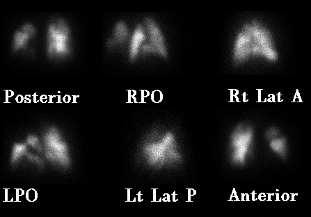


What are pathophysiologic sequelae of PE?
1. Anatomic obstruction of pulmonary arteries... although pulmonary infarction is typically avoided since lungs are also supplied by bronchial arteries.
2. Platelets cause the release of serotonin and vasoactive substances resulting in elevated pulmonary vascular resistance and V/Q mismatch.
3. RV afterload increases, leading to RV dilatation, ischemia, and right sided heart failure.


What are chronic sequelae of DVT and PE?
Post-thrombotic syndrome (chronic leg pain/swelling)
Chronic thromboembolic pulmonary hypertension


What are classic risk factors for DVT?
Virchow's triad - stasis, injury, and hypercoagulability

Also advanced age, obesity, pregnancy + postpartum period, malignancy, reduced mobility, polycythemia vera, smoking, central venous catheters, HITS, antiphospholipid antibodies, lupus anticoagulant
Medications - chemotherapy, oral contraceptives (3X risk), hormone-replacement therapy (2x risk), tamoxifen
Acutely-ill, hospitalized, medical patients have a risk of 15%


What specific populations are at high risk for DVT?
Total hip and knee replacement (risk of >50% without prophylaxis)
Cancer surgery
Major trauma
Spinal cord injury


What are genetic disorders that predispose to risk of venous thromboembolism?
Protein C and S deficiency
Antithrombin deficiency
Factor V Leiden
Prothrombin mutation (g20210a)
Hyperhomocyteinemia (C677T mutation)
Dysfibrinogenemia
Plasminogen deficiency


What isthe most common genetic risk factor for thrombophilia?
Factor V Leiden. This causes resistance to activated protein C


What are signs and symptoms of lower extremity DVT?
Leg pain, tenderness, warmth, or swelling

Homan's sign - calf pain with passive dorsiflexion at the ankle


What are signs and symptoms of PE?
dyspnea (73%)
tachypnea (70%)
pleuritic chest pain (66%)
rales / crackles (51%)
cough (37%)
Tachycardia (30%)
Leg swelling (28%) or pain (26%)
Fourth hearth sound (24%)
Loud P2 (23%)
Hemoptysis (13%)
Palpitations (11%)
JVD, systolic murmur, right sided gallop
syncope
hypotension
hypoxemia
PEA/cardiac arrest

Cardiogenic shock is diagnosed in 5% of acute PE.
While hypoxia and an Aa gradient is common, a normal ABG is seen in up to 20% of PE.


What are ECG findings of PE?
Tachycardia
V1-V4 T wave inversion
S1-Q3-T3 pattern
Right bundle branch block
P-wave pulmonale
Right axis deviation

These are nonspecific and are more often seen with very large PE


What are abnormal findings on CXR attributed to PE?
Westermark's sign - focal peripheral oligemia caused by PE obstruction
Hampton's hump - peripheral wedge seen above diaphragm
Palla's sign - enlarged right descending PA


How does D-dimer testing help with a diagnosis of PE?
This is a sensitive (96-98%) but very non-specific test.
It must be considered together with clinical suspicion and is better for patients presenting to the emergency room than for general surgical postop patients.
If the DDimer is negative in patients with a low-to-moderate pretest probability, imaging is not useful since PE is very unlikely.
If the patient has a high pretest probability, don't bother with checking a DDimer and go straight to imaging.


What imaging strategies are used for diagnosis of PE?

Spiral CT-angiography (90% sensitivity, 95% specificity)
Ventilation/Perfusion Scanning - limitation: frequently non-diagnostic
Pulmonary arteriogram
Venous duplex for DVT
Echocardiography for hemodynamically significant PE


Use caution when Cr>1.5 with CTA
Avoid V-Q scanning if there is associated cardiopulmonary disease noted on CXR


What are treatment stategies for DVT?
Outpatient management with low-molecular weight heparin transitioning to warfarin with a goal INR of 2-3 for 3-6 months

Pregnant patients can't take warfarin and must be kept on a LMWH...


What are treatment strategies for PE?
PE should be managed by inpatient hospitalization.
Parenteral unfractionated heparin (therapeutic PTT), low-molecular weight heparin, or fondaparinux for at least 5 days while starting warfarin - bridging therapeutic INR of 2-3 for 2 consecutive days, continuing treatment for 3-6 months.

Factor VII - measured by the INR - has a half life of 6hrs... however thrombin (factor II) depletion takes 5 days...


How is unfractionated heparin administered?
It is bolused 60-80 U/kg, followed by a continuous infusion of 18 U/kg/hr. The infusion is titrated to a therapeutic PTT (60-80 seconds) by checking the PTT level every 6 hrs.


What are benefits of LMWH?
These have more predictible pharmacokinetics and greater bioavailability than unfractionated heparins, allowing subcutaneous administration once or twice per day.

Therapeutic dosing for enoxaparin is 1mg/kg SC BID or 1.5mg/kg SC QD


How can you monitor if levels of LMWH are therapeutic?
Check anti-factor Xa levels

This is not as convenient as a PTT, but consider this in morbidly obese or pregnant patients, or if there have been issues with renal function.


What is fondaparinux?
A synthetic pentasaccharide that inhibits Factor Xa
Structurally, it looks like the antithrombin-binding portion of heparin. It is administered once daily, subcutaneously.

This should not be used in patients with severe renal insufficiency.


What if the patient has heparin-induced thrombocytopenia with thombosis?
The patient should be treated with direct thrombin inhibitors (argatroban or lepirudin). Warfarin treatment should be held until the disease process is controlled and the platelet count has normalized to avoid thrombotic complitcations and warfarin-induced skin necrosis.

Lepirudin is excreted by the kidneys and argatroban is metabolized by liver.


What is the risk of death from recurrent thromboembolism in patients treated for DVT?
0.4%


What is the risk of death from recurrent thromboembolism in patients treated for PE?
1.5%


What are indications for a vena caval filter?
Contraindications to anticoagulation
Major bleeding complications during anticoagulation
Recurrent DVT or PE while receiving adequate therapy

Retrievable filters can often be inserted... although often times they are forgotten to be removed!
The tide is beginning to turn against prophylactically placing IVC filters, but they may be recommended in select trauma patients.


When is thrombolytic therapy considered for management of PE?
If the PE is severe enough to cause cardiogenic shock or right ventricular dysfunction

There is no hard data to answer this question... surgical embolectomy may also be considered in certain cases of massive PE when conservative measures fail.


What is the overall mortality rate for PE?
15% at 3 months


What is the optimal duration of therapy for a DVT or PE
If it is secondary to a reversible risk factor, including the postoperative state, 3-6 months should be sufficient.
If it is idiopathic or secondary to a documented hypercoagulable state, 6-12 months can be considered.
Patients with a DVT/PE secondary to cancer should be treated until their cancer has resolved, with a LMWH for whe first 3-6 months.
Patients with recurrent thromboembolisms may require indefinite therapy.


Late... SG

References:
Goldhaber SZ. Pulmonary Embolism. NEJM 339; 1998: 93-104.
Tapson VF. Acute Pulmonary Embolism. NEJM 358; 2008: 1037-1052.
Cameron's Current Surgical Therapy (9th ed.)

Wednesday, July 9, 2008

Acute Calculous Cholecystitis


A recent edition of NEJM has a nice review on this common surgical topic...

What proportion of patients with gallstones become symptomatic?

1-4% per year


What proportion of patients with symptomatic cholelithiasis develop cholecystitis?
20%


What is gangrenous cholecystitis?
When the gallbladder wall undergoes necrosis and gangrene.


What is emphysematous cholecystitis?
When gas is visible on imaging in the wall or lumen of the gallbladder... this is indicative of superinfection with gas-forming organisms and may lead to perforation without urgent intervention.


What is Mirizzi's Syndrome?
Obstruction of the common bile duct as a result of extrinsic compression of a stone within the gallbladder or cystic duct.


What is Murphy's Sign?
Arrest of inspiration while palpating the gallbladder during a deep breath.


What are ultrasonographic findings of acute calculous cholecystitis?
Gallstones
Gallbladder wall thickening, greater than 5mm
Pericholecystic fluid
Positive ultrasonographic Murphy's sign

It's also important to assess for choledocholithiasis by evaluating for a common bile duct dilated greater than 7mm or frank CBD stones.


The PPV of stones + Murphy's sign is 92%.
The PPV of stones + GBW thickening is 95%.
The NPV of no stones, and a normal GBW and no Murphy's sign is 95%.


What does a HIDA scan involve?
Hepatic scintigraphy uses an IV injection of technetium-labelled iminodiacetic acid analogues. These are excreted by the liver into bile, and allows visualization of the gallbladder within 30 minutes. An absence of filling of the gallbladder after 60 minutes indicates cystic duct obstruction and is 80-90% sensitive for cholecystitis.

Morphine can improve the specificity of the test by increasing resistance at the sphincter of Oddi. Overall, HIDA has greater specificity and accuracy in comparison to ultrasound, but is usually reserved when the diagnosis of cholecystitis is uncertain.


What is a 'rim sign' on HIDA?
A pericholecystic blush that is seen in 30% of patients with acute cholecystitis and 60% of patients with gangrenous cholecystitis.


What are the Tokyo guidelines?
Diagnostic criteria for Acute Cholecystitis:
Presence of one local sign/symptom, one systemic sign, and any confirmatory finding on an imaging test...
Local signs and symptoms: Murphy's sign, RUQ pain or tenderness, RUQ mass
Systemic signs: Fever, leukocytosis, elevated CRP

According to the Tokyo guidelines, acute cholecystitis can be subdivided into three grades of severity...


What are criteria for Grade 1 - Mild Cholecystitis?
No organ dysfunction
Does not meet criteria for a more severe grade

Early laparoscopic cholecystectomy is recommended.


What are criteria for Grade 2 - Moderate Cholecystitis?
The presence of any of the following:
WBC greater than 18,000
Palpable, tender RUQ mass
Duration greater than 72hrs
Marked local inflammation including pericholecystic abscess, hepatic abscess, gangrenous or emphysematous cholecystitis, or biliary peritonitis

Early or delayed laparoscopic cholecystectomy may be considered depending on the scenario and the surgeon's expertise.


What are criteria for Grade 3 - Severe Cholecystitis?
Organ system dysfunction - hypotension requiring pressors, mental status changes, respiratory insufficiency with PaO2/FiO2 less than 300, oliguria, Cr greater than 2.0, INR greater than 1.5, platelet count less than 100k

Management with antibiotics and percutaneous cholecystostomy tube may be considered, reserving surgery for treatment failures.


What is the optimal timing of cholecystectomy?
For most patients, early cholecystectomy may be favored. 15-20% who had their procedures delayed after the initial attack subsided require intervention before their planned lap chole.

In several studies/meta-analyses, there were no differences in operative time or conversion rates between early and delayed lap chole. However, bile duct injuries in general may be more common in the setting of acute cholecystitis. Postoperative bile duct leaks were more common with early intervention (3%) compared to delayed (0%).


What are the rates of conversion from laparoscopic to open during cholecystectomy for acute cholecystitis?
5-30%

These are greater in acute cholecystitis compared to uncomplicated cholelithiasis, but there is no difference between early and delayed intervention.


When should antibiotics be administered in the setting of cholecystitis?
Per the IDSA, a second-generation cephalosporin or a fluoroquinolone and metronidazole shold be administered in the following scenarios:
WBC greater than 12,500 or Temperature greater than 38.5C AND
Radiographic findings of cholecystitis
Also in elderly, diabetics, or immunocompromised

Patients undergoing cholecystectomy will also get a prophylactic perioperative dose, even if they don't require empiric treatment.


When should percutanous cholecystostomy tubes be used?
Patients with Grade 3/Severe cholecystitis
Septic shock
Poor surgical candidates

Delayed cholecystectomy may be considered at a later time.


Late... SG

References:
Strasberg SM. Acute Calculous Cholecystitis. NEJM 2008; 358: 2804-2811.

Sunday, September 16, 2007

Square Knot Tutorial

It's been awhile... here's a sweet tutorial on knot tying.


Late... SG

Wednesday, May 30, 2007

Rectal Prolapse


What is the difference between procidentia and rectal mucosal prolapse?

Procidentia is a complete, full-thickness, rectal prolapse.

Procidentia can be differentiated by its circumferential, concentric folding pattern. Mucosal prolapse manifests with deep grooves with a radial pattern.

What patient population typically presents with rectal prolapse?
Older women

What studies may be useful in evaluation of rectal prolapse and disorders in defecation?
Colonic transit studies
Anorectal manometry
Pudendal nerve latency studies
Electromyography
Cinedefecography

What studies should be performed prior to operative intervention?
Colonoscopy or air-contrast barium enema

The colon should be assessed for neoplasms and diverticular disease.

What are the two primary surgical approaches to manage rectal prolapse?
1. Abdominal
2. Perineal

The abdominal approaches have lower recurrence rates, while perineal approaches have lower operative morbidity. There are also laparoscopic modifications that can potentially decrease surgical stress on the patient.

What procedures utilize the abdominal approach?
1. Rectopexy (suture or prosthetic)
2. Rectopexy with resection of redundant sigmoid

What procedures utilize the perineal approach?
1. The Delorme procedure
2. The Altemeier procedure
3. The Tiersch procedure

What is the Moschowitz repair?
Transabdominal reduction of a perineal hernia with closure of the cul-de-sac

What is the Ripstein repair?
A transabdominal rectopexy using an anterior mesh sling

What is the Wells repair?
A transabdominal rectopexy using a posterior mesh suspension

What is the Delorme procedure?
A transperineal mucosectomy

This is commonly performed for mucosal prolapse and is essentially a mucosal sleeve resection with plication of the muscularis.

What is the Altemeier procedure?
A transperineal rectosigmoidectomy

This involves eversion of redundant rectum with a full-thickness resection and anastomosis.

What is the Tiersch procedure?
An anal encirclement procedure that was originally described using a silver wire to encircle the external sphincter within the ischiorectal space, creating an outlet obstruction.

This is considered a palliative procedure, and has been done under local anesthesia using subcutaneously placed polypropylene mesh.

Late...SG

Sources:
Schwartz' Principles of Surgery, 8th ed.
Mastery of Surgery, 5th ed.

Monday, April 16, 2007

Electrosurgery


Who first demonstrated the principles of electrosurgery?

Alex d'Arsonval, a French physicist


Who first produced an electrosurgical unit designed for human use?

William Bovie, a Harvard biophysicist


Who popularized the use of "the Bovie"?

Harvey Cushing, MD, the father of modern neurosurgery


Cushing is reknown for developing surgical techniques that reduced the mortality rate of brain surgery from 90% to under 10%.


What is Cushing's Disease?

Secondary hypercortisolism, from a pituitary tumor leading to ACTH-hypersecretion.


Cushing's Syndrome is a more general term, referring to any form of symptomatic glucocorticoid excess, whether from an adrenocortical tumor, or exogenous steroids.


What prestigious award did Cushing receive in his academic career?

The Pulitzer Prize, for his biography "A Life of Sir William Osler"


What is Ohm's Law?

Voltage = Current * Resistance


What is Impedance?

A measure of resistance to flow when using alternating current (AC)


A high cellular water content results in a low tissue impedance.


What is Alternating Current?

Current that cycles in direction at a specific frequency... this comes from a typical US wall outlet at rates of 60Hz.


Electrocautery utilizes direct current (i.e. battery-powered hand-held units), while electrosurgical units utilize alternating current.


What radiofrequencies cause neuromuscular stimulation?

Up to 100kHz


What radiofrequencies cause a generation of heat?

200kHz - 5MHz


What is the Joule-Lenz Law?

Heat dissipated = (Current-density)squared * Resistance * time


Current-density is current divided by cross-sectional area.
Thus, heat generated with electrosurgery is inversely proportional to the area of the electrode and proportional to the time of contact and the impedance of the tissue.


What is monopolar current?

Electricity flowing from a single electrode to the patient.


Monopolar units typically utilize a pencil-type probe that is hand operated. Laparoscopic monopolar cautery may be operated by a foot pedal.


What is a dispersive electrode?

A ground or grounding pad.

Why is a ground pad placed on the patient when using monopolar cautery?

It allows completion of the electric circuit through the patient back to the electrosurgical unit.


Does the size of the ground pad matter?

By being large, it disperses current over a greater surface area. If it were small (i.e. detached/poor contact), it could cause generation of enough heat to create a local burn.


What is bipolar current?

Electricity flows in a closed circuit between two closely placed electrodes (i.e. bipolar forceps or scissors). Current flow and injury to tissue is limited to the region between the electrodes.


Bipolar units typically allow the use of a lower voltage waveform than monopolar units to acheive hemostasis.


What are common modes used by monopolar electrosurgical units?

1. Cutting
2. Coagulation
3. Blend


What are the properties of a cutting waveform?

It uses a continuous (pure sine-wave), low voltage, high-frequency waveform (500kHz) that concentrates energy on a small area.
Instantaneous heating (100 degrees C) results in cellular evaporation at contact, with minimal damage to surrounding tissues.


What are properties of a coagulation waveform?

It uses a pulsed, higher-voltage waveform that allows for dispersal of energy deeper into tissue.
Heating is less (37- 60 degrees C), causing tissue dessication and protein denaturing, allowing for sealing of blood vessels.


What is fulgaration?

Surface coagulation, accomplished by a non-contact method of electrocautery.


What are the properties of a blend mode?

This is a combination of cutting and coagulation waveforms. The higher the blend, the higher the coagulation effects.


What happens to tissue impedance during coagulation?

As water evaporates and tissue dessicates, the tissue impedance increases, leading to increased coagulative effect.


This can also increase the potential of arcing of the current to a lower impedance tissue, leading to iatrogenic injury.


Remember, current flows in the path of least resistance.


What are special hazards seen with laparoscopic electrosurgery?

1. Insulation failure
2. Direct coupling
3. Capacitor coupling


Injury can occur to non-target tissue out of view of the surgical team!


What is insulation failure?

When a defect in the covering of the active electrode allows current to contact non-target tissue.


What is direct coupling?

When the tip of the active electrode comes into contact with another metal instrument in the surgical field.


What is capacitor coupling?

When stray current from an electrode develops by a storage of charge. This can be minimized by activating the active electrode only when it is in contact with target tissues and limiting the amount of time on the coagulation setting.


This can sometimes occur with devices that contain both plastic and metal, allowing for insulation of charge by the instrument or trocar.


For a resource that has some pretty decent illustrations, consider this online course at Valleylab.

Late... SG

Sources:
O'Leary's Physiologic Basis of Surgery, 3rd ed.
Jones CM, Pierre KB, Nicoud IB, Stain SC, Melvin WV. Electrosurgery. Curr Surg 2006; 63: 458-463.

Monday, April 9, 2007

Aphorisms and Quotations


Here's a book for any quote enthusiast:

Aphorisms & Quotations for the Surgeon. Edited by Moshe Schein. tfm Publishing, Ltd. : Shrewsbury, UK; 2003.


For those of you who have been looking for a convenient resource to pepper your next powerpoint presentation with a breath of surgical history, this is the book for you. Even better if you are looking for a little inspiration at the end of the day. In addition to instilling the text with witticisms of his own, Moshe Schein has organized the numerous quotes into 94 different subject headings, ranging from the ubiquitous organ systems, to topics such as academics, dogma, greatness, love, money, politics, gimmicks, fame, ethics, empathy, and errors.


Here are a few:


There are 4 degrees of intra-operative hemorrhage:
1. "Why did I get involved in this operation?"
2. "Why did I become a surgeon?"
3. "Why did I study to become a doctor?"
4. "Why was I born?"
-Alexander A Artemiev


When I am carrying out a big, unusual, or difficult operation, I never plan anything later that day.
-Francis D. Moore


It is a most gratifying sign of the rapid progress of our time that our best textbooks become antiquated so quickly.
-Theodor Billroth


I would like to see the day when somebody would be appointed surgeon somewhere who had no hands, for the operative part is the least part of the work.
-Harvey W. Cushing


Medical examinations: they are no tests of the man. They are only tests for his memory for facts. They tell us nothing of his judgement, tact, energy, enthusiasm, idealism, reason, observation, temperament, disposition, honesty, loyalty, courage, truthfulness, or intelligence. Memory of facts means little. The other things mean nearly all.
-J. Chalmers Da Costa


The pleasure of a physician is little, the gratitude of patients is rare, and even rarer is material reward, but these things will never deter the student who feels the call within him.
-Theodor Billroth


You must always be students, learning and unlearning till your life's end, and if, gentlemen, you are not prepared to follow your profession in this spirit, I implore you to leave its ranks and betake yourself to some third-class trade.
-Joseph Lister


Late... SG

Saturday, April 7, 2007

Pancreatic Transplantation


Who discovered insulin?

Banting and Best (1921)


Sir Frederick Banting of the University of Toronto was awarded the Nobel Prize for Medicine/Physiology along with John MacCleod in 1923. He initially refused to accept the award until his research assistant Charles Best's work was acknowledged, but ultimately ended up sharing his portion of the award with Best.


What are the secondary complications of diabetes mellitus?

Retinopathy
Nephropathy
Neuropathy
Enteropathy
Vasculopathy


Diabetes is the leading cause of kidney failure, blindness, nontraumatic amputation, and impotence.


Exogenous insulin can prevent the acute metabolic complications and decrease the secondary complications of diabetes, but cannot maintain a homeostatic environment.


What is the benefit of pancreatic transplantation?

It can establish normoglycemia and insulin independence and can halt the progression of secondary complications of diabetes


What is the drawback of pancreatic transplantation?

Immunosuppression


What are the methods of pancreatic transplantation?

1. PAK - pancreas after kidney transplantation
2. SPK - simultaneous pancreatic/kidney transplantation
3. PTA - pancreas transplant alone


Often PAK and SPK is performed because immunosuppression is already required for a kidney transplant.
PTA may be performed for nonuremic diabetics with a poor quality of life from ketoacidosis, labile glycemic control, or progression of secondary complications of diabetes.


When was the first pancreatic transplant performed?

1966 as an SPK. This was three years after the first kidney transplant.


What techniques are used in the drainage of the pancreas?

1. Enteric drainage (requires creatinine monitoring, for PAK or SPK)
2. Bladder drainage (requires monitoring of urine amylase)


Enteric drainage was performed in the 1970's but was superceded by ureteral, then bladder drainage. Bladder drainage became common in the 1980's, but in the 1990's enteric drainage became repopularized. Enteric drainage has been thought to be more physiologic, but requires a kidney transplant to monitor for rejection.


What are elements of the preoperative evaluation for pancreatic transplantation?

1. Assessment of degree of kidney dysfunction
2. Evaluation of secondary complications of diabetes
3. Coronary Angiography
4. Evaluation of peripheral arterial disease and iliac vessel patency
5. Rule out Malignancy
6. Rule out Infection


Why is coronary angiography often routinely performed?

Because severe diabetics are at risk for coronary arterial disease without angina.


What are the options based on kidney function?

1. PTA if Cr<2.0mg/dL with minimal proteinuria
2. PAK or SPK with moderate kidney insufficiency


What are the donor possibilities for kidney/pancreas transplants?

1. Deceased-donor SPK
2. Living-donor kidney transplant followed by deceased-donor PAK
3. SPLK: Simultaneous deceased-donor pancreas and living-donor kidney transplant
4. Living-donor SPK


What are the elements of preparation of the pancreas graft?

1. Removal of the spleen and excess duodenum
2. Ligation of the vessels at the root of the mesentery.
3. Reconstruction of the arterial system of the graft (Y-graft)


What is a pancreatic Y-graft reconstruction?

The donor SMA and splenic artery are connected using a reversed segment of donor iliac artery.


This creates a common arterial inflow channel for the pancreatic graft


What is the outflow of the pancreatic graft?

Portal vein


What are the components of the pancreatic transplant?

1. Heterotopic placement of pancreatic graft (right iliac fossa or mid-abdomen)
2. Arterial anastomosis: Y-graft to common iliac artery or distal aorta
3. Venous anastomosis: donor portal vein to recipient iliac vein (systemic) or recipient SMV (portal)
4. Drainage procedure: duodenocystostomy or duodenoenterostomy (i.e. Roux en Y)


What are complications associated with bladder drainage?

Dehydration, chronic refractory metabolic acidosis, recurrent UTI, hematuria, bladder calculi, urethritis, urinary leaks


10-20% with bladder drainage are converted to enteric drainage secondary to complications


When is bladder drainage commonly used?

With PTA (allows for rejection monitoring via urinary amylase levels), or when the kidney and pancreas are from different donors.


What are signs of rejection after pancreatic transplants?

-increased creatinine (kidney/pancreas transplants)
-decreased urinary amylase (bladder drainage)
-increased serum amylase
-increased serum glucose


What are other complications of pancreatic transplantation?

1. Infections (10%)
2. Thrombosis (6%)
3. Hemorrhage (<1% of graft loss)
4. Pancreatitis
5. Urologic complications (see bladder drainage)


What are the reported graft survival rates after pancreatic transplantation?
SPK - 90% at 1yr
PAK - 85% at 1yr
PTA - 75% at 1yr


What is a pancreatic islet cell transplant?

Cells are extracted from the islets of Langerhans of donor pancreas and injected into a diabetic patient's portal vein.


This potentially avoids a major surgical procedure, but the patient will still need long-term immunosuppression (use is still often limited to patients needing kidney transplants).


What are problems with Islet cell transplantation?

Islet cell rejection is difficult to monitor and diagnose
Complications such as hepatic abscesses, bacteremia, and portal hypertension can occur
Poor results (<5% insulin-independence at 1yr)


Late... SG

Source:
Schwartz' Principles of Surgery, 8th ed.